BRAF V600E mutation in two distinct meningeal melanocytomas associated with a nevus of Ota
Fecha
2014Resumen
Meningeal melanocytomas are rare tumors of the CNS that develop from melanocytes that are present in leptomeninges, with differing pigmented appearance. They generally occur in the posterior fossa and the spinal cord. This lesion may manifest at any age, but most patients are in the fifth decade of life.1,2 Occasionally, these tumors appear in a complex neurocutaneous grouping with other melanocytic lesions. The nevus of Ota (oculodermal melanocytosis) is a blue hyperpigmented dermal lesion that affects the trigeminal dermatome. The association of a meningeal melanocytoma with an ipsilateral nevus of Ota is extremely rare; to our knowledge, only eight cases have been reported in the literature to date.3,4 In these cases, the melanocytomas were located in the supratentorial area.
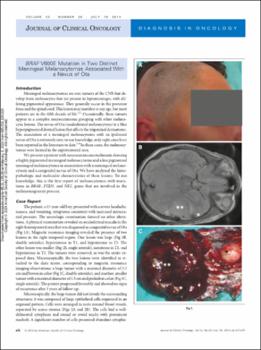
We present a patient with neurocutaneous melanosis showing a highly pigmented meningeal melanocytoma and a less pigmented meningeal melanocytoma in association with a meningeal melanocytosis and a congenital nevus of Ota. We have analyzed the histopathologic and molecular characteristics of these lesions. To our knowledge, this is the first report of melanocytomas with mutations in BRAF, PTEN, and NF2, genes that are involved in the melanomagenesis process.


